Cervical lymph node superresolution imaging
Development of ultrasound localization microscopy for the visualization of the microvascular anatomy of cervical lymph nodes
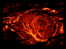
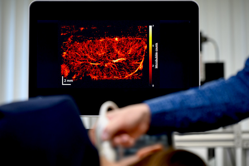
The prognosis of patients with squamous cell carcinoma of the head and neck region is largely determined by the tumor involvement of the cervical lymph nodes. To date, non-invasive assessment of the cervical lymph nodes using clinical imaging techniques has not been solved satisfactorily due to their insufficient sensitivity and specificity. Therefore, possible metastasis must be ruled out at least by biopsy, but usually by removing and analyzing individual lymph nodes. However, the unnecessary removal of healthy lymph nodes is stressful for patients and possibly even unfavorable due to the important role of the lymph nodes in the induction of the anti-tumor immune response. Accordingly, there is a need for better non-invasive diagnostics of metastasized lymph nodes. One starting point for diagnostic imaging is the visualization of the vascular supply to the lymph node, which is altered early on by metastasis. However, it has not yet been possible to visualize the relevant very small vessels with low flow velocities using any clinical imaging method. This project therefore aims to investigate the potential of ultrasound localization microscopy (ULM), which was co-developed by the applicants, for the early detection of lymph node metastases. This method is based on contrast-enhanced sonography, in which microbubbles are injected into the bloodstream. The basic idea of the method is that these microbubbles can be localized individually in video sequences with high spatial accuracy and then tracked. As the localization accuracy is better by a factor of 5-10 than the resolution of the ultrasound image, this results in a high-resolution image of the vessels through which microbubbles travelled. This allows smaller vessels to be visualized than is possible with previous sonographic methods, e.g. low-flow Doppler imaging. The project now aims to clarify which parts of the vasculature can be imaged with ULM and whether quantitative parameters derived from the ULM images, such as vessel densities, flow velocities and vessel structure parameters, show differences between healthy and metastasized lymph nodes. In the study, contrast-enhanced sonography is carried out in several parallel sectional planes in order to enable nearly complete coverage of the lymph node. The ULM images are calculated, and both known and new quantitative parameters are calculated to characterize the vasculature. Micro-computed tomography of complete lymph nodes embedded in paraffin and histological sections spatially assigned to the ultrasound images are used as a control method. This makes it possible to check the generated vascular images and the quantitative parameters using a reference method. Changes in the parameters between healthy and tumor tissue are examined for both methods. As a result, a decision can be made if a larger-scale clinical trial to validate non-invasive lymph node staging is indicated.


